Ongoing Research
Master’s Thesis: The evolution of arthropod trachea during the water-land transition
Understanding how new body parts arise is one of evolutionary biology’s greatest mysteries. Insects and millipedes are different lineages that both moved from water to land. Both groups have air breathing tubes called tracheae, but a lack of ‘trachea-like’ structures in their common ancestor has led to the belief that these similar structures must have evolved independently. My research challenges this idea by going beyond the fossils and using modern molecular techniques to uncover their evolutionary history. I will use in-situ HCR to visualize the expression patterns of tracheal and leg patterning genes in millipede embryos. If the trachea in both animals develop in the same location, using the same genes, they likely evolved from the same ancestral structure. This would suggest that both groups evolved tracheae from the same ancestral gill at different times. This would redefine how we think new animal structures arise, suggesting it is easier for developmental and genetic systems to remodel old structures beyond recognition than to invent new ones.
Developing a lab-tractable myriapod model
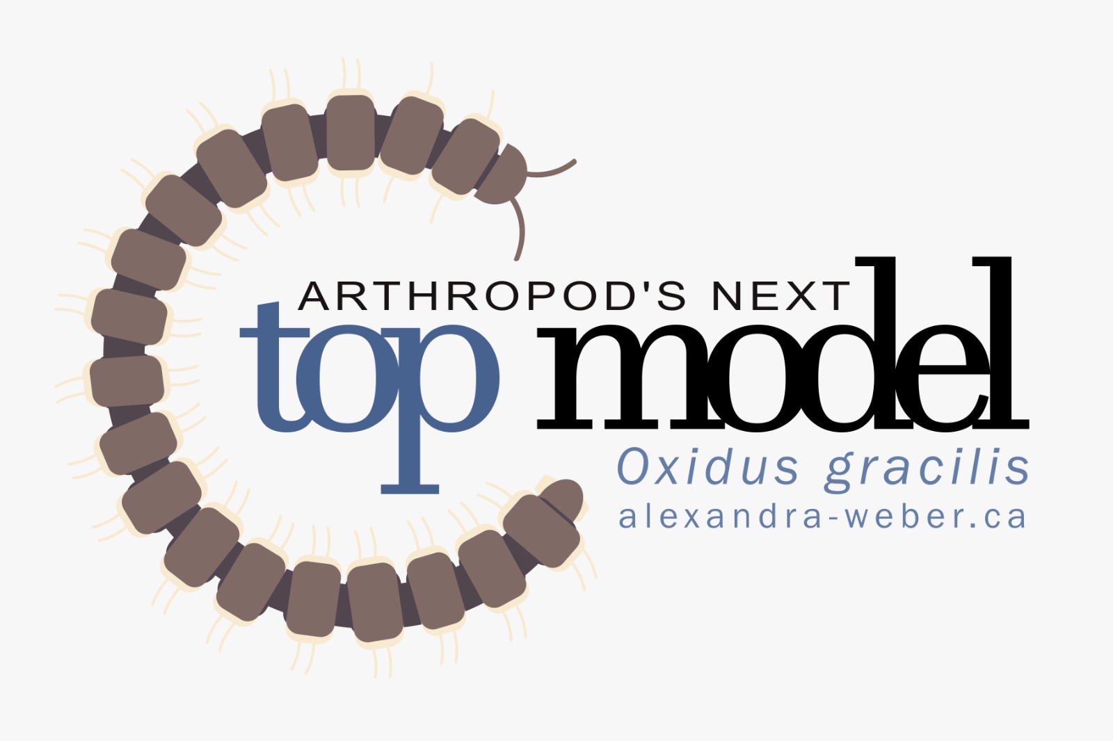
There are 4 major extant groups within Arthropoda: Chelicerata, Myriapoda, Crustacea and Insecta. Of these groups, Myriapoda (millipedes and centipedes) is the only not to have an established lab tractable model. However, there is keen scientific interest due to their key phylogenetic position between Chelicerates and Pancrustacea (Crustaceans and Insects), and unique traits like cyanide-based defensive glands, distinctive neural patterning, tracheal systems, and extreme take on Arthropoda’s characteristic segmented body plan. I am developing Oxidus gracilis as the first lab-tractable myriapod model with a sequenced and annotated transcriptome, complete lab life cycle, and embryo staging manual. In contrast to other myriapod species examined to date, *Oxidus is an ideal model system: 1) it thrives in lab environments; 2) breeds year-round; 3) produces clutches of >200 eggs; 4) has a short generation time of ~4 months; and 5) is readily available worldwide. Pest control is the biggest issue, with nematodes, mold and phoretic mites all posing threats to the health of the animals. I am currently working on publishing the lab rearing protocol.
Past Projects

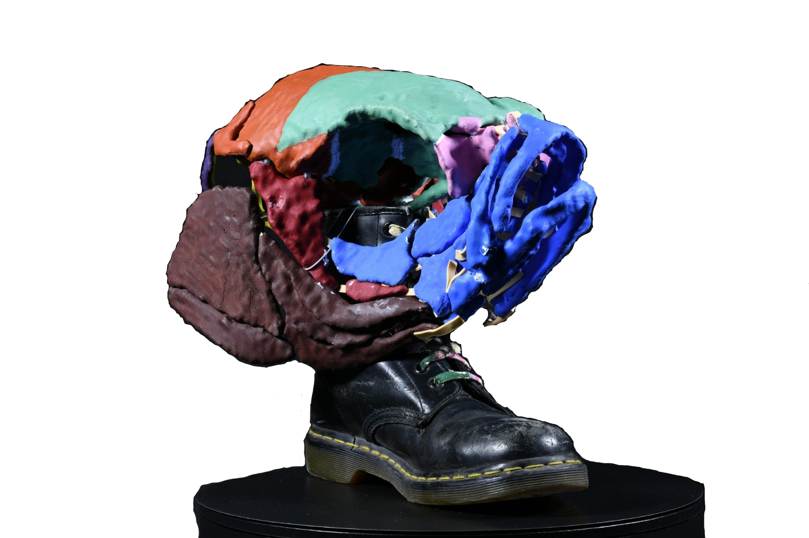
Undergraduate Thesis: Comparative Skull Anatomy of Adult Zebrafish (Danio rerio) and Functionally Jawless nkx3.2−/− Mutant.”
The evolution of articulating jaws was a major innovation in vertebrate history, enabling more efficient feeding and expanding ecological opportunities. During my undergrad I researched how jaw joint fixation alters skull development and function using nkx3.2 knockout zebrafish. These “jawless” mutants survive to adulthood, offering a rare opportunity to examine the plastic remodelling that occurs in the absence of an articulating jaw. Although nkx3.2-/- mutant phenotype is not atavistic, their skull shape and kinematics resemble Paleozoic jawless vertebrates such as anaspids and thelodonts. This convergence implies that integration and modularity in the vertebrate skull constrain plastic responses in functionally predictable ways. These findings position nkx3.2 mutants as a model for understanding how jaw articulation shapes cranial evolution, and how developmental plasticity may have shaped the jawed vertebrate phenotype before they became genetically fixed.
The kinematic models created for this project are currently on display at the Canadian Museum of Nature in the new exhibit “Life onto Land: The Devonian”.
Astrobiology
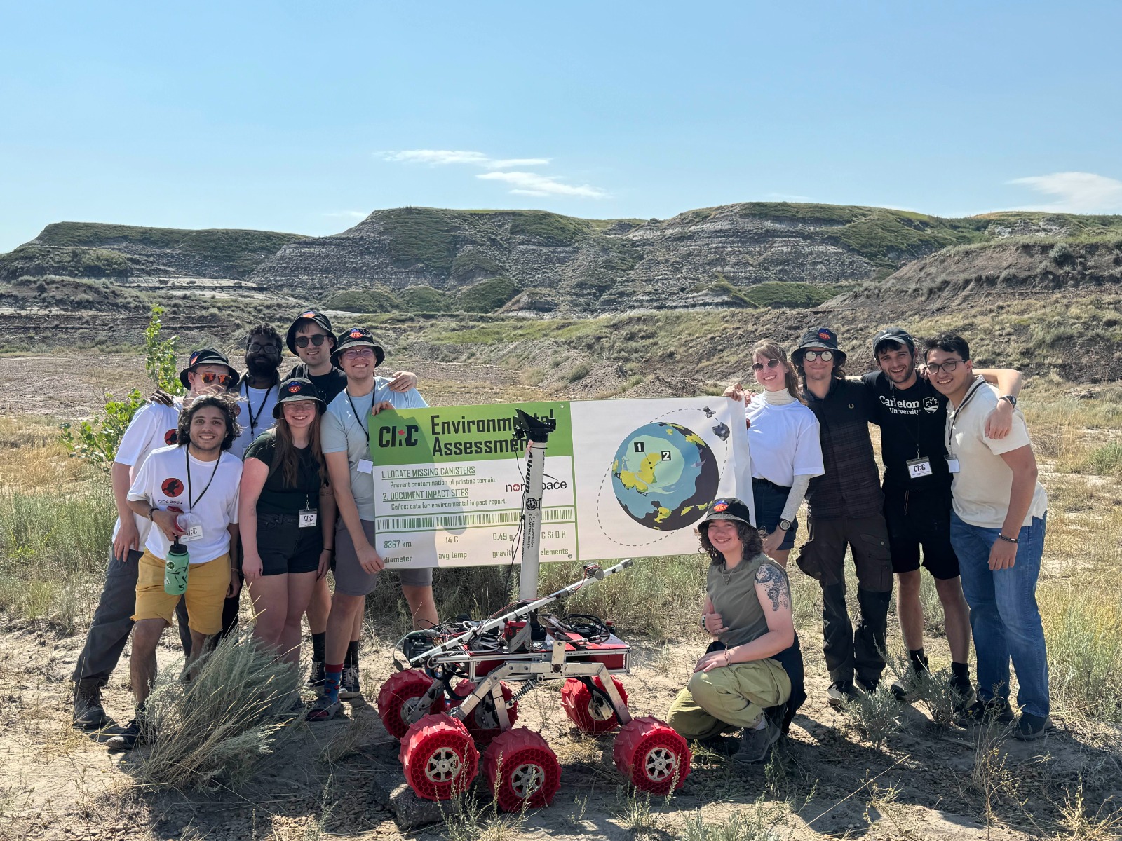
Science Lead – Carleton Planetary Robotics Team
The Carleton Planetary Robotics Team is an engineering design team that builds and develops a Mars rover and competes at international university competitions. As the science sub-team lead, I oversaw the overall design of the life detection and environmental analysis capabilities onboard our rover. My other responsibilities include managing the Science sub-team members and creating a welcoming and inclusive environment. I also made it my personal mission to teach engineers about the scientific method, and the philosophy of science. During my tenure in the role, we achieved the highest team score for the science environmental analysis mission at the Canadian International Rover Competition and qualified for the University Rover Challenge, helping secure our spot as one of the top teams in the world.
Symmetry Breaking in Snake Embryos: How and Why They Coil
I began this project in the second year of my undergrad degree. It is currently in review.
Abstract: Snake embryos are often tightly coiled, but the biological basis for this developmental trait remains a puzzle. To understand how and why they coil, we first investigated directions of coiling within and across 39 snake species. In early developmental stages, coiling orientation appears to be nearly fixed right-handed across multiple species. We present evidence that accelerated somitogenesis outpaces gastrointestinal extension at these stages, causing the embryos to coil around the delayed visceral tissues. To allow this, the body axis and the gut are physically separate, where the latter forms a pillar-like structure from the stomach to cloaca. The dextral directional bias in these early embryos is likely passively determined by the left-sided yolk mass. Coiling becomes looser and more inconsistent later in development when the embryos acquire mobility through maturation of axial musculoskeletal tissues and with a sufficiently extended gut. These consilient lines of observations reveal in embryonic coiling a passive process where its initiation and direction are each a response to spatial conditions of snake embryogenesis. As these parameters conflict each other at some developmental stages (e.g., the offset growth curves between gut and body axis), snakes apparently solve some of the many challenges to patterning their startlingly elongate bodies by taking advantage of what could otherwise be discordant features of the embryonic growth.
Survey of Spotted Wing Drosophila Populations in Southern Ontario
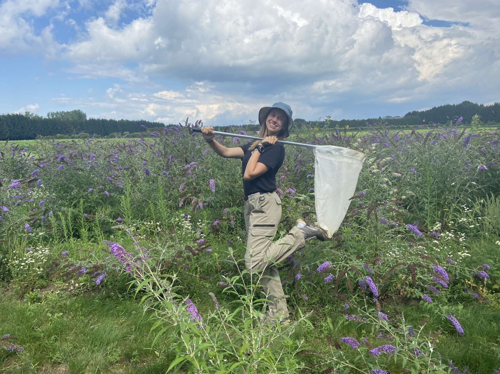
In 2022 I completed a co-op work term at Agriculture and Agri-Food Canada in the Molecular Diagnostics and Biological Control of Invasive Insect Pests in Agriculture lab. My main project was a survey of spotted wing drosophila (Drosophila suzukii) populations across Southern Ontario, and for the detection of possible parasitoids. D.suzukii is an invasive agricultural pest that, unlike other fruit-fly species, lays eggs within ripe soft skinned berries making them unsellable. Spotted wing drosophila has multiple natural parasitoids in its native habitat, that keep the populations at a manageable level. This project aimed to monitor the population of both D.suzukii and their parasitoids and inform the controlled release of these parasitoids into high population areas. I also assisted with was a survey of the population distribution of the three main parasitoids of the pest Lygus lineolaris. This project was a follow up to a previous controlled release of Peristenus digoneutis. The objectives of this project were to see if the parasitoid was able to establish in Ontario, and if it is more commonly found in Lygus populations then the generalist parasitoid native to Ontario.